

/ News, Research / Angelika Jacobs
New molecules against hard-to-treat tumors (Hutter Lab)
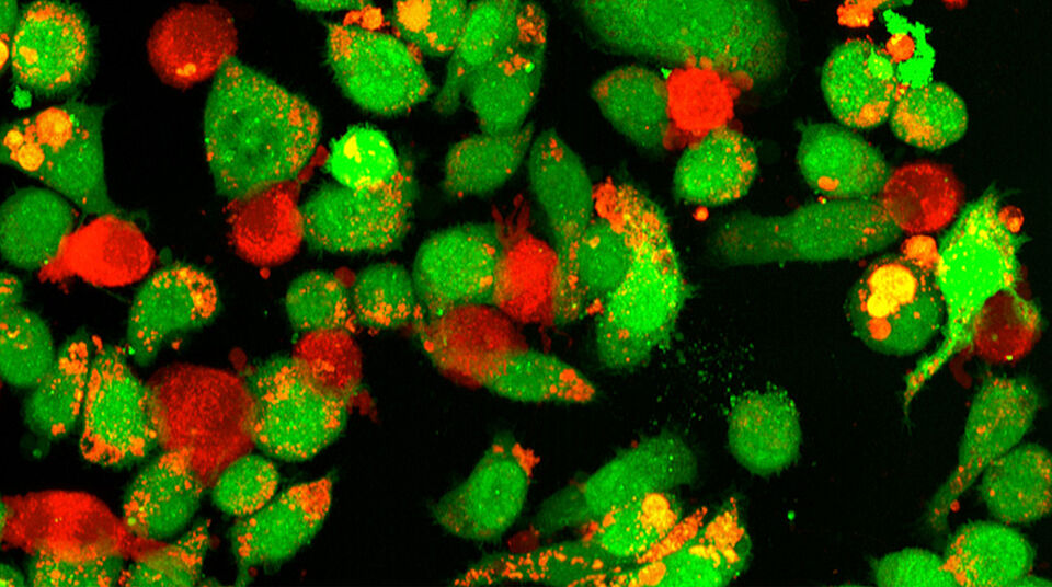
Researchers from Cambridge and Basel have developed small molecules that could make cancer immunotherapies more effective. They work in two ways: They prompt macrophages to engulf cancer cells and alter the surrounding tissue, reducing its ability to suppress immune attacks on the tumor.
“Cold tumors” is the term experts use for cancers that shield themselves so effectively from the immune system that therapies often have little effect. New therapeutic approaches are therefore needed.
Researchers are increasingly turning their attention to the tissue surrounding tumors. Cancer reshapes this environment to its own advantage. One molecule, PD-L1, plays a key role: It acts like a stop signal for the immune system’s T cells, preventing them from attacking the tumor.
“But PD-L1 is found not only on the cancer cells themselves but also on certain immune cells in the tumor microenvironment known as macrophages or phagocytic cells,” says Professor Gregor Hutter of the University of Basel and University Hospital Basel. “Cancer effectively co-opts these immune cells to suppress T-cell responses.”
“Immunotherapy has focused almost entirely on T cells, but the myeloid cells that dominate most solid tumors have been the missing piece,” says Professor Gonçalo Bernardes of the University of Cambridge. “Our work shows they can be reprogrammed to attack the tumor rather than shield it.”
A molecular cable with an “eat” signal
The research teams led by Bernardes and Hutter have now developed small molecules that activate macrophages in the immediate vicinity of the tumor, causing them to engulf cancer cells. At the same time, the molecules weaken the tumor’s defenses against the immune system. The two teams report their findings in the journal Cancer Research.
The so-called “Phagocytic Synapse Enhancers,” or PSEs for short, act like a sophisticated molecular cable with an added function. They connect phagocytic cells directly to cancer cells, triggering the phagocytes to engulf and break down their targets.
To do this, each PSE molecule has two functional ends. One docks onto said PD-L1, the other end carries the molecule tuftsin that binds to and activates phagocytic cells. “We are essentially creating a molecular bridge between the tumor cells and the macrophages and giving the macrophages the signal to attack,” explains Dr. Valerio Sabatino, first author of the study and a research associate in Gregor Hutter’s group. Sabatino conducted the study in Cambridge and Basel with support from a Postdoc.Mobility grant from the Swiss National Science Foundation.
Tumor microenvironment turns against cancer
In addition, PSE molecules cause macrophages to internalize and break down PD-L1 from their own surface. This weakens the stop signal that restrains T-cell activity. “We are reprogramming the tumor microenvironment from one that supports the tumor to one that works against it,” explains Valerio Sabatino.
The researchers tested their approach in cell cultures and in zebrafish and mouse models of different tumor types. A modified PSE variant with a longer residence time in the body was particularly effective: It slowed tumor growth more than the comparator treatments and extended survival.
Customizable system
The researchers show that PSEs could serve as a modular system: Individual components can be swapped out to tailor the molecules to different tumor types or target structures.
The scientists now plan to further develop the PSEs for potential clinical use. “Our long-term vision is to design CAR T cells for cancer therapy that not only attack the tumor directly but also release PSE molecules there to activate macrophages as well,” says Gregor Hutter. However, much more research will be needed before PSEs can be tested in clinical trials.