Molecular Imaging . Ultrasound . Microbubbles . Atherosclerosis . Vascular Inflammation
Cardiovascular Molecular Imaging
Ultrasound molecular imaging in cardiovascular disease
Ultrasound molecular imaging
Developments in hardware, computing power and image processing have contributed to impressive improvements in medical imaging over the last decades with depiction of the human body with ever increasing resolution. However, early disease processes are often characterized by changes in cellular phenotype and will remain out of range for conventional imaging. To overcome this limitation, molecular imaging that uses contrast media to image biological processes at a cellular level has been developed for all major medical imaging modalities, and it is thought that molecular imaging will in the future allow to detect diseases earlier, lead to better understanding of pathophysiology and will be an integral part of personalized medicine. In our laboratory, we are developing targeted contrast agents for ultrasound molecular imaging. Microbubble ultrasound contrast agents have been in use in the clinical field for some years for blood pool and perfusion imaging. With a size range of 1–4μm, microbubbles circulate freely throughout the microcirculation. Being composed of a gas core and a monolayer lipid shell, microbubbles re-radiate sound waves efficiently and can be depicted using clinical ultrasound equipment. For ultrasound molecular imaging, microbubbles have been functionalized for molecular imaging by anchoring antibodies or other ligands to the microbubble surface (Fig. 1). Upon injection, these functionalized microbubbles accumulate on the vascular endothelium expressing a particular marker in diseased tissue, and can be imaged non-invasively with ultrasound.
Ultrasound molecular imaging in atherosclerosis
Risk assessment for atherosclerotic cardiovascular events currently relies on clinical risk factors. This approach places a large proportion of individuals in an inter-mediate risk category, where the value of interventions to reduce the risk for events is uncertain. Therefore, tools to better assess the risk in these patients are needed. Noninvasive imaging of molecular events associated with atherosclerotic disease may serve this purpose. Previous studies have shown that contrast enhanced ultrasound molecular imaging using microbubble contrast agents directed against vascular cell adhesion molecule 1 (VCAM-1), which is involved in inflammatory processes in atherosclerosis, is feasible in murine disease models. However, the ultrasound contrast agents used in these studies are not suitable for clinical translation and there is a need for the development of microbubbles employing clinically translatable strategies for conjugation of targeting moieties, and targeting ligands that can readily be used in the clinical field. Therefore, recent work in our laboratory has concentrated on the development of contrast agents that will allow translation of this non-invasive imaging technique into the clinical field. Nanobodies are small antibody fragments (10–15kDa) derived from heavy-chain-only antibodies. They are attractive for applications in molecular imaging, as they are highly specific, non-immunogenic and thus offer the potential for clinical translation. We have shown that MBs bearing nanobodies with cross reactivity for murine and human VCAM-1 can be not only used to image various stages of murine atherosclerosis, but that imaging of VCAM-1 expression on human carotid thrombendarterectomy specimens is also possible using an in-vitro flow chamber setup (Fig. 2). Likewise, Designed Ankyrin Repeat Proteins (DARPins) are potential candidates for clinical ultrasound molecular imaging given their easy production and selection, high affinity and low immunogenicity, and we have examined their use as ligands for targeting of VCAM-1. For this purpose, ribosome display was used to select >400 DARPin binders targeted against murine VCAM-1. Subsequently, flow cytometry and flow chamber assays were used to select 5 top candidate binders that were assessed in-vivo. Using these top candidates, assessment of murine hindlimb inflammation was possible using ultrasound molecular imaging.
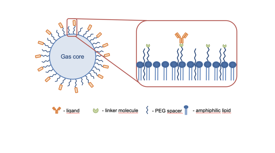
Fig. 1: Microbubble contrast agent used for ultrasound molecular imaging. Microbubbles consist of a lipid monolayer shell and a heavy weight gas core. The shell is functionalized with a specific ligand bound to the surface through linker molecules.
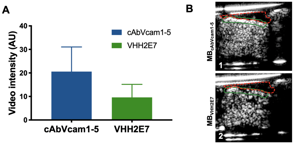
Fig. 2: (A) Background-subtracted ultrasound molecular imaging signal intensity on the luminal endothelial surface of human endarterectomy specimens (n=7) for microbubbles targeted to VCAM-1 (MBcAbVcam1-5) or control microbubbles (MBVHH2E7). Data is mean ± SD, MBcAbVcam1-5 showed increased retention (*p=0.0156) as compared to MBVHH2E7. (B) Example of ultrasound molecular imaging showing high signal on the plaque surface for MBcAbVcam1-5 (panel 1) and low signal for MBVHH2E7 (panel 2). The red color outline shows the human thromboendarterectomy specimen, the green color outline attached microbubbles at the tissue-fluid interface.