Angiogenesis . Bioreactors for 3D Culture and Physical Stimulation . In vitro 3D Cardiac Models . Small Diameter Vascular Grafts .
Cardiac Surgery and Engineering
3D Engineered tissues as angiogenic therapeutic approach and as functional cardiac models
The main goal of our group is to investigate repair therapies based on engineered tissues (patches) to induce safe and efficacious angiogenesis and to rescue hibernating myocardium in a chronic ischemic myocardium. The three-dimensional (3D) bioreactor culture of heterogenous Stromal Vascular Fraction (SVF) cells aims to standardize the production of engineered patches (Fig.1). SVF as a cell population is capable of releasing a broad secretome range comprising angiogenic and prosurvival factors. Further aims are to develop in vitro functional cardiac models to investigate processes of myocardial repair and regeneration.
Research is funded by the Swiss National Science Foundation, Swiss Heart Foundation, the University Hospital of Basel (USB), and the University of Basel (Unibas).
Patches engineered by cells with a high angiogenic and repair potential
In this approach, we use human adipose tissue-derived SVF cells as a heterogeneous cell population with a high angiogenic potential thanks to the presence of numerous endothelial and mural progenitors (collaboration with A. Scherberich, Tissue Engineering Group, Department of Biomedicine, Unibas). We demonstrated that perfusion-based bioreactor culture supported the maintenance of endothelial and mural cells as compared to static culture, thereby accelerating whole construct vascularization and support of cell survival upon implantation in a subcutaneous nude rat model (Fig. 1–2). Moreover, medium conditioned during 3D perfusion- based culture of SVF was showed to partially rescue damaged cardiomyocyte function during monolayer culture under sever hypoxic condition (<1% of oxygen) (Mytsyk and Isu, 2018). An ongoing in vivo diseased study aims to investigate the repair potential of SVF-perfused patches in nude rat model of cardiac ischemia (in collaboration with the Department of Cardiac Surgery, USB).
3D functional cardiac models
Our angiogenic engineered tissues might also affect cardiac repair and regeneration by influencing cardiomyocyte maturation and functionality while increasing progenitor cell recruitment. Therefore, we aim to generate a 3D functional cardiac models as a tool to investigate the interactions of SVF cells’ secretome and cardiomyocytes. We hypothesize that the recapitulation of the proper physiological conditions, mimicking the native tissue environment, enhanced the cardiomyocyte maturation, 3D organization and functionality. Culture medium perfusion systems were employed to mimic the highly dense capillary network present in the myocardium to ensure the cardiomyocyte survival in vitro (Marsano, et al. 2010; Maidhof, et al. 2010; Cerino, et al. 2016). Mechanical stimulation (collaboration with the Politecnico of Milano, Italy and Politecnico of Torino, Italy) was employed to greatly promote rat neonatal cardiomyocytes or human induced pluripotent stem cellderived cardiomyocyte maturation and contractility both at the micro- and macro- scale (Marsano, et al. 2016, Massai and Pisani, et al. 2020). Microfluidic culture systems harnessing mechanical stimulation was recently generated to recapitulate some of key steps of a scar formation (Occhetta, Isu, et al. 2019).
A novel 24-multi-well bioreactor (Patent number: EP19165964) was also developed to culture mm-scale engineered cardiac constructs in a highly controlled environment under multi-physical stimulations (isotonic and auxotonic loads combined with electrical stimulation).
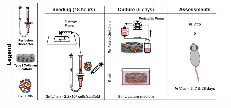
Fig. 1: Scheme of the SVF-based patch study. Summary of the main steps to generate SVF cell-based engineered tissues (patches). SVF cells were seeded on a collagen-based scaffolds under direct alternated perfusion for 18 hours followed by 5 days of culture either under direct uni-directional perfusion or in static condition. The resulted patches were assessed in vitro and in vivo (in subcutaneous pockets of nude rats).
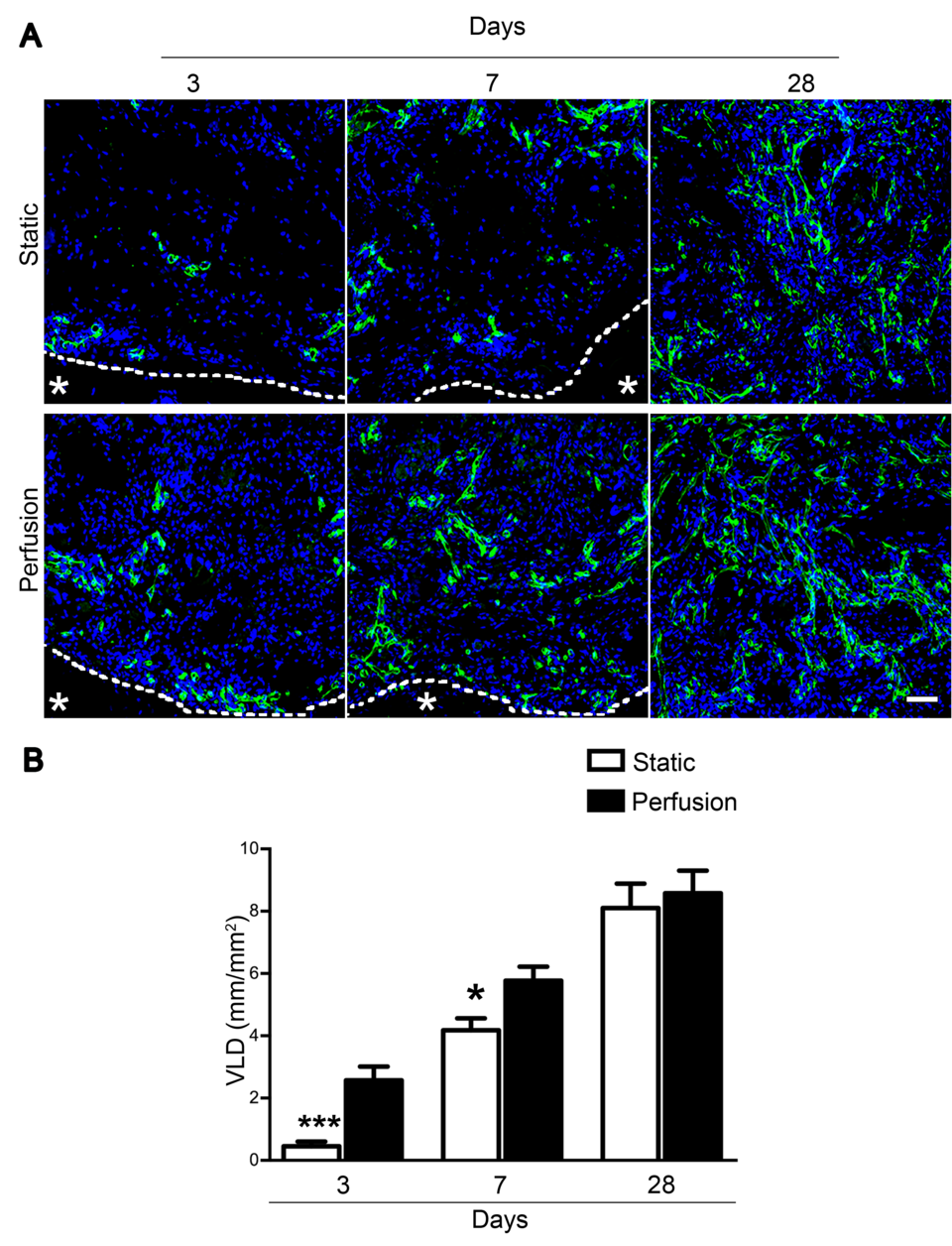
Fig. 2: Perfusion accelerated in vivo vascularization of engineered tissues. Engineered patches generated either under perfusion or in static culture were analysed after 3, 7, and 28 days in vivo. (A) Representative immunofluorescence images stained for endothelial cells (CD31; green). Dashed lines outline the border between the patch and rat tissue (identified by the *). Scale bar= 50 μm. (B) VLD quantification normalized over the analysed area.