Subchondral Bone Plate . Distribution . Mechanical Properties . Osteochondral Unit Biomechanics . Functional adaptation
Musculoskeletal Research
Form-Function-Relationship in the locomotor apparatus and application in the living as diagnostic tools or for monitoring the outcome of joint surgery.
CT-Osteoabsorptiometry (CT-OAM) – a new investigation technique in the field of mummy research
The objective of the current study was to investigate the applicability of CT-OAM on mummies for the load analysis of joints. In order to clarify whether apparent malpositions of the spinal column existed during lifetime or occurred post-mortem, we evaluated the long-term loading patterns within the endplates of 8 mummies. The implementation of CT-OAM on mummies for load analysis of joints was feasible. The mineral density distribution within the endplates was not homogenous but followed distinct distribution patterns. The vertebra columns without malposition showed an almost even circular allocation of the density maxima within thoracic endplates, the spines with kyphosis displayed a concentration of the density maxima in the ventral area, and the spines with scoliosis exhibited a predominant localization of the density maxima on the concave side. The examined endplates showed characteristic reproducible density patterns consistent with the long-term loading conditions. Pathological load distributions can be visualized with help of CT-OAM before macroscopical changes appear and can therefore help to solve paleopathological and paleoarchaeological questions.
CT-calculometry (CT-CM): advanced NCCT post-processing to investigate urinary calculi
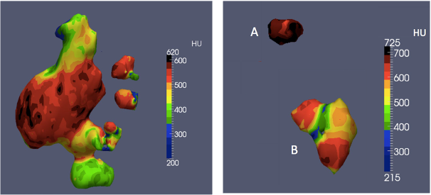
This study aimed at evaluating the potential of CT-calculometry (CT-CM) as a novel method to determine mineralisation, composition, homogeneity and volume of urinary calculi based on preoperative non-contrast-enhanced computed tomography (NCCT) scans. CT-CM was performed in preoperative NCCTs of 25 patients treated for upper tract urinary calculi by ureterorenoscopy or percutaneous nephrolithotomy. Absolute mineralisation values were achieved by use of quantitative CT-osteoabsorptiometry and compared to Fourier infrared spectroscopy as a reference for stone composition. Homogeneity was assessed by advanced software-based NCCT post-processing and visualised by using a maximum intensity projection algorithm. Volumetric measurement was performed by software-based threedimensional reconstruction. CT-CM was feasible in all of the 25 NCCTs. Absolute mineralisation values calculated by quantitative CT-OAM might be used to identify the most frequent stone types. High levels of inhomogeneity could be detected even in pure component stones. Volumetric measurement could be performed with minimal efort. CT-CM is based on advanced NCCT postprocessing software and represents a novel and promising approach to determine mineralisation, composition, homogeneity, and volume of urinary calculi based on preoperative NCCT. CT-CM could provide valuable information to predict outcome of diferent stone treatment methods.
Adaption of the bony-microstructure of the human glenoid cavity due to long-term biomechanical loading
Structural arrangements of the bony microstructure of a joint through adaptational processes are thought to be determined by the biomechanical demands. Pursuing this theory of “form follows the biomechanical function”, the load distribution of the glenoid cavity, as it is mirrored in its mineralization pattern, should also have an impact on the trabecular network below. To prove this hypothesis, we analyzed the mineral distribution and the distribution of the architectural parameters of the trabecular network below. Our findings clearly show an inhomogeneous but regular and reproducible mineral distribution. Regarding the trabecular network below, the distribution of the analyzed parameters also revealed an inhomogeneous distribution with a regular pattern in correlation with the biomechanical impact. With increasing depth, the trabecular network administers the expression of each structural parameter given that the strain energy becomes increasingly evenly distributed and changes from a high degree of differentiation just beneath the subchondral bone plate to a more equal distribution within the deeper areas. Thus, the biomechanical situation of a joint directly influences the bony formation of the subchondral bone plate and trabecular network below.
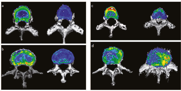
Fig. 1: Examples of the mineralization distribution, a densitograms of thoracic endplates in vertebral columns without malposition, on the left: Mummy Lausanne 2, on the right: L-25–14, b densitograms of lumbar endplates in vertebral columns without malposition, on the left: Mummy Lausanne 2, on the right: L-25–14 , c densitograms of thoracic end-plates in vertebral columns with kyphosis, on the left: Mummy Barfuesser, on the right: L-32–13 d densitograms of lumbar endplates in vertebral columns with scoliosis, on the left: Mummy Baroness von Kniestaett, on the right: L-05–15.
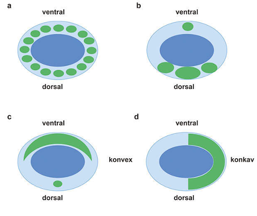
Fig. 2: Schematic illustration of the found mineralization patterns, dark-blue presents the little mineralized center, light-blue the higher mineralized marginal zone and green the localization of the density maxima a normal thoracic endplate, b normal lumbar endplate, c kyphotic endplate, d scoliotic endplate.